
Translate this page into:
An unusual configuration of anterior capsular phimosis in an elderly asian patient

*Corresponding author: Benjamin Chong Ming Chang, Department of Ophthalmology and Visual Sciences, Khoo Teck Puat Hospital, Singapore. chang.benjamin.cm@ktph.com.sg
-
Received: ,
Accepted: ,
How to cite this article: Pandiyan PS, Chang BC. An unusual configuration of anterior capsular phimosis in an elderly Asian patient. J Ophthalmic Res Pract. 2024;2:42-3. doi: 10.25259/JORP_8_2024
Anterior capsule contraction syndrome (ACCS) or anterior capsular phimosis refers to the fibrosis of the continuous curvilinear capsulorhexis following cataract surgery. It tends to occur in patients with conditions of zonular weakness, intraocular inflammation, uveitis, and pseudoexfoliation.[1] The possible pathophysiology of ACCS involves remaining metaplastic lens epithelial cells on the capsular bag differentiating and forming fibrosis.[2] Complications of ACCS include reduction in visual acuity (VA), intraocular lens (IOL) decentration or tilt and traction on the zonules, resulting in pseudophacodonesis, IOL dislocation, and retinal detachment.[3] Neodymium: Yttrium-aluminium-garnet (Nd: YAG) laser radial anterior capsulotomy has been safely and effectively used to enlarge the opening area in ACCS.[4]
An 88-year-old systemically stable patient underwent right eye phacoemulsification and IOL implantation. The fellow eye had been operated 7 months earlier with no complications. Pertinent perioperative findings were that he was on tamsulosin, an alpha-1 blocker; he had small pupils requiring iris hooks, and he was noted to have loose zonules.
Post-operative day 1 findings were unremarkable. At postoperative week 5, he was found to have developed ACCS with VA of 6/15. Notably, there was a central pillar of fibrosis with a clear region temporal to the pillar [Figure 1a]. It was uncertain if the clear region represented a physical defect in the anterior capsule. The clinical dilemma of whether to disrupt the central pillar emerged as there was concern of IOL dislocation if it was disrupted. The decision was made not to completely disrupt the pillar and to take a more conservative approach with small radial incisions to the margins of the capsular phimosis and for partial radial nicks at the central pillar.
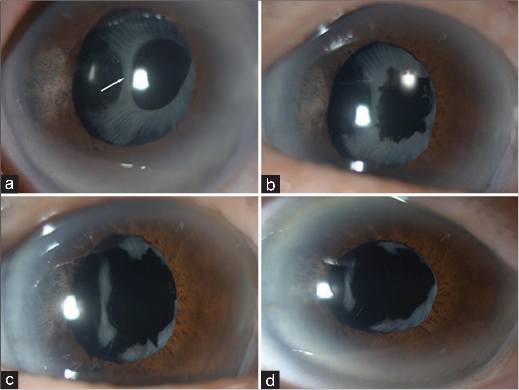
- An 88-year-old male with weak zonules, small pupil requiring iris hooks and on an alpha-1 blocker (tamsulosin) – post-cataract surgery with an odd configuration of anterior capsule contraction syndrome. Slit-lamp photos showing (a) central pillar of fibrosis (white arrow) and clear region of capsule temporally, (b) after first Neodymium: Yttrium-aluminium-garnet (Nd: YAG) laser radial anterior capsulotomy, (c) after second Nd: YAG laser radial anterior capsulotomy, and (d) after third Nd: YAG laser radial anterior capsulotomy showing that the previously noted central pillar has shifted temporally out of visual axis.
The patient was reviewed 1 week later with improvement in VA and reduced glare but the central pillar remained in the visual axis [Figure 1b]. A second Nd: YAG laser radial anterior capsulotomy was performed extending the nicks above, below, and at the pillar with the result 1 month later as per Figure 1c.
The patient reported improved vision with best-corrected VA of 6/7.5 but was still symptomatic with glare when looking at bright lights. A third laser Nd: YAG capsulotomy was performed with the intention of shifting the central pillar temporally with the result 2 weeks later as per Figure 1d. The patient, then, reported satisfactory vision with VA of 6/6.7 and alleviation of the previous symptoms. At post-operative month 3, the patient had no complications, including that of IOL dislocation. Nd: YAG laser radial anterior capsulotomy was performed safely in this patient with an unusual configuration of anterior capsular phimosis.
Ethical approval
The Institutional Review Board has waived the ethical approval for this study.
Declaration of patient consent
The authors certify that they have obtained all appropriate patient consent.
Conflicts of interest
There are no conflicts of interest.
Use of artificial intelligence (AI)-assisted technology for manuscript preparation
The authors confirm that there was no use of artificial intelligence (AI)-assisted technology for assisting in the writing or editing of the manuscript and no images were manipulated using AI.
Financial support and sponsorship
Nil.
References
- Postoperative occlusion of visual axis with fibrous membrane in the presence of anterior capsular phimosis in a patient with pseudoexfoliation syndrome: A case report. BMC Ophthalmol. 2016;16:213.
- [CrossRef] [PubMed] [Google Scholar]
- Lens cell populations studied in human donor capsular bags with implanted intraocular lenses. Invest Ophthalmol Vis Sci. 2000;41:1130-41.
- [Google Scholar]
- Anterior capsule opacification: International ophthalmology clinics. Available from: https://journals.lww.com/internatophthalmology/citation/2001/07000/anterior_capsule_opacification.4.aspx [Last accessed on 2024 Feb 25]
- [Google Scholar]
- Long-term follow-up of neodymium: YAG laser anterior capsulotomy for the treatment of anterior capsular phimosis. J Int Med Res. 2018;46:3692-7.
- [CrossRef] [PubMed] [Google Scholar]


